



Fibular hemimelia is a rare congenital anomaly characterized by partial or complete failure of the external bone (fibula) under the knee to form during development in the womb, causing severe shortness of the leg and outward bending at the ankle. Thanks to modern pediatric orthopedic approaches, today, instead of amputation, the limb can be successfully preserved with reconstructive surgeries that stabilize the ankle and computer-assisted leg lengthening surgeries. Prof. Dr. With his advanced surgical experience in the treatment of congenital fibula deficiency and complex limb shortness, Cengiz Çabukoğlu enables children to walk into the future with balanced, healthy and independent steps on their own legs.
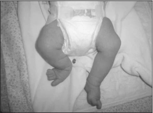
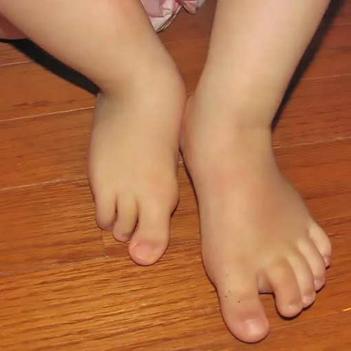
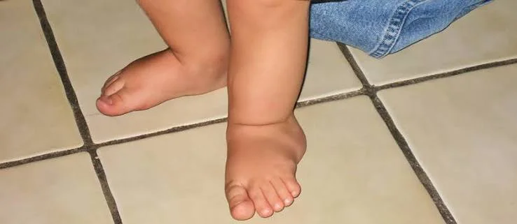
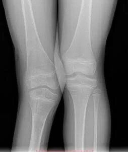
One of the most complex disorders that disrupt muscle, bone and joint development in childhood is congenital anomalies, that is, congenital limb deficiencies, which begin in the womb. Fibular hemimelia, one of these rare and specific anomalies, is a condition in which the outer one (fibula/peroneal bone) of the two bones located between the knee and ankle is partially or completely not formed at birth. Lack of this bone, which provides the external support column of the leg and the external balance of the ankle; It causes severe congenital leg shortness complaints in children, dangerous outward bending of the ankle, and deficiencies in the toes. This biomechanical problem, which directly affects the developmental process, should be addressed with a multifaceted treatment schedule by a pediatric orthopedist specialized in this field, starting from the moment of birth. The clinical picture in babies born with fibular hemimelia may vary greatly depending on the degree of bone deficiency, from mild shortness to advanced stages in which the ankle is completely dysfunctional. When the baby grows and tries to stand up and participate in the walking cycle, the length difference between the two legs and the instability (looseness and insecurity) in the ankle make it impossible for him to make a balanced step. In these children, shortness of the thigh bone (femur) and looseness of the kneecap ligaments often accompany the process. For this reason, orthopedic radiographs and detailed clinical examinations in early infancy are of vital importance in order to accurately analyze the functional potential of the limb and create a surgical schedule appropriate to the child's growth rate. In the past years, when it came to the treatment of advanced fibular hemimelia, cutting the leg below the knee (amputation) considering that it would not be functional and directing the child to a prosthesis at an early age was considered one of the most common solutions. However, today's modern

 Çerez Tercihleri