



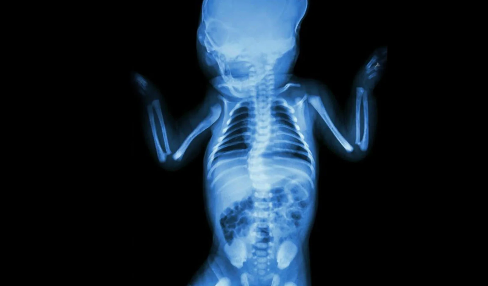
Other Congenital Anomalies
Category Pediatric Orthopedics
Congenital muscular and skeletal anomalies are babies being born with structural defects in their hands, feet or spine. It is successfully treated with pediatric orthopedic follow-up.
What is the problem?
Congenital anomalies in children are structural deformities of the musculoskeletal system that occur during fetal development due to genetic, environmental, or unknown factors. Under pediatric orthopedics, the category of "Other Congenital Anomalies" encompasses a wide spectrum of common and rare conditions beyond developmental hip dysplasia or major bone deficiencies. These include skeletal conditions such as webbed digits (syndactyly), extra digits (polydactyly), radial clubhand (radial longitudinal deficiency), congenital knee dislocation, and twisted neck (torticollis).
What are the symptoms?
Congenital musculoskeletal anomalies present at birth with distinct signs depending on the specific condition:
Hand and Foot Deformities: Presence of extra fingers or toes (polydactyly) or webbed/fused digits (syndactyly).
Wryneck (Torticollis): The baby's head consistently tilts to one side while the chin points toward the opposite shoulder, often accompanied by a firm, palpable mass in the neck muscle.
Limb Deformities: Joint bending in an abnormal direction or extreme hyperextension of the knee backwards (congenital knee dislocation).
Joint Rigidities: Multiple joints throughout the body remaining stiff, immobile, and fixed in a bent position from birth (Arthrogryposis Multiplex Congenita).
What causes it?
While there is no single cause underlying congenital anomalies, errors during early embryonic development play a central role:
Genetic and Hereditary Factors: Familial gene mutations or chromosomal abnormalities (such as Down syndrome or specific skeletal dysplasias).
Intrauterine Mechanical Pressure: Restricted space in the womb due to low amniotic fluid levels (oligohydramnios) or multiple pregnancies, which places physical stress on the developing joints.
Environmental Factors: Maternal infections during the first trimester of pregnancy, unprescribed medication use, or exposure to harmful chemicals.
How is it diagnosed?
Most congenital anomalies can be diagnosed early thanks to modern medical technology:
Detailed Prenatal Ultrasound: Level II ultrasound scans performed during pregnancy can detect major structural abnormalities in the hands, feet, and limbs (such as polydactyly or severe limb deformities).
Clinical Physical Examination: A comprehensive systemic examination conducted by a pediatric orthopedist immediately after birth evaluates joint range of motion and digit anatomy.
Imaging Modalities (X-Rays and MRI): Plain X-rays are used to evaluate bone formation and alignment, while Magnetic Resonance Imaging (MRI) provides detailed visualization of soft tissues, cartilage, and ligaments.
Treatment methods
Treatment approaches are tailored to the individual based on the specific type of anomaly, the child's overall health, and the degree of functional impairment:
Conservative (Non-Surgical) Treatments: Particularly effective for torticollis and mild joint stiffness, early-intervention stretching exercises, pediatric physical therapy, and serial casting yield high success rates.
Orthotics and Splinting: Custom-fabricated splints and orthopedic braces are utilized to hold joints in proper anatomical alignment and support healthy development.
Is surgery required?
Yes. Surgical intervention is required in cases that do not improve with conservative methods or directly impede the child's growth, hand function, or ability to walk.
For instance, webbed digits (syndactyly) are surgically separated to enable independent finger movement, while extra digits (polydactyly) are excised to restore normal hand or foot anatomy. In cases of congenital knee dislocation or severe joint contractures (such as in arthrogryposis), tendon release, ligament repair, and bone realignment surgeries are performed.
Recovery process
The recovery process varies depending on the extent of the surgical intervention. Following soft tissue procedures, such as digit separation or excision, wound healing typically takes 2 to 3 weeks, during which protective dressings are applied. For larger operations involving bone and joint reconstruction, the affected limb may remain in a cast for several weeks.
Post-surgery, a structured pediatric physical therapy program combined with regular pediatric orthopedic follow-ups is essential to ensure long-term surgical success, restore full joint mobility, and build muscle strength.
Frequently asked questions
Surgical treatment of congenital anomalies of the fingers and toes is usually planned before the child begins to develop fine motor skills, that is, between the ages of 6 months and 1.5 years (18 months). Operations performed in the early period are the ideal time for the child to perceive his hand normally and for the healthy direction of bone development.
Yes, more than 90% of torticollis cases diagnosed in the first 6 months recover completely without the need for surgery, thanks to regular stretching exercises at home and professional pediatric physical therapy under the supervision of a pediatric orthopedist. In resistant cases that do not improve after the age of 1, a short muscle relaxation surgery is required.
Although it varies depending on the severity of the anomaly and the region it affects, thanks to today's pediatric orthopedic surgery and microsurgery methods, most of the anomalies are completely corrected or the child is enabled to live his life independently, aesthetically and functionally like his peers.
Video
Appointment and information for Other Congenital Anomalies
Contact us with your questions about the treatment process; we will arrange the most suitable appointment together.
 Çerez Tercihleri