

deformity
Category Pediatric Orthopedics
Limb deformities in children are bone curvatures and lengths in the arms or legs. It can be corrected non-surgically or surgically with early pediatric orthopedic follow-up.
What is the problem?
Limb deformity in children refers to any alteration in the normal anatomical structure, alignment, or length of the bones in the arms, legs, or spine. Frequently encountered in pediatric orthopedics, these conditions often manifest as inward or outward leg curvature (knock-knees or bowlegs), bone twisting (rotational deformities), and differences in leg length (leg length discrepancy).
Because children’s growth plates are actively developing, these structural deformities can progress as the child grows—or, with timely and proper intervention, be successfully corrected and controlled.
What are the symptoms?
Bone and joint deformities in children typically present with the following signs:
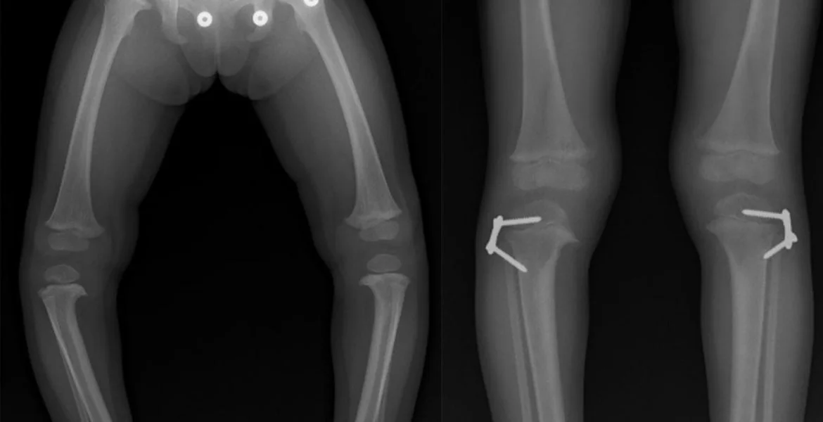
Knock-Knees or Bowlegs: Knees that angle in and touch each other (Genu Valgum / X-legs) or bow outward leaving a wide space between them (Genu Varum / O-legs).
In-Toeing or Out-Toeing: Walking with feet pointed noticeably inward or outward.
Visible Length Discrepancies: A noticeable difference in length between the left and right arms or legs.
Gait and Stamina Issues: Rapid fatigue during walking or running, frequent loss of balance, tripping, or a noticeable limp.
Postural Abnormalities: Spinal alignment issues such as side-to-side curvature (scoliosis) or an exaggerated rounded upper back (kyphosis).
What causes it?
The underlying causes of childhood deformities can be either congenital (present at birth) or acquired (developmental):
Congenital Factors: In-utero positioning during pregnancy, genetic syndromes, or congenital conditions that affect bone development.
Growth Plate Injuries: Damage to the growth plates (physeal plates) caused by bone fractures or infections during childhood.
Nutritional and Metabolic Disorders: Conditions that weaken the bone structure, such as Rickets (caused by severe Vitamin D deficiency) or Blount’s disease.
Neuromuscular Disorders: Diseases affecting the nerve and muscle systems—such as Cerebral Palsy—which can alter bone alignment over time.
How is it diagnosed?
Diagnosing a deformity begins with a comprehensive pediatric orthopedic evaluation. The specialist analyzes the child's gait and precisely measures joint range of motion and leg lengths. To reach a definitive diagnosis and calculate the exact degree of curvature, the following imaging methods are used:
Full-Length Standing Radiography (Orthoradiogram): A specialized X-ray technique that captures the entire leg from hip to ankle on a single image, allowing millimeter-precise calculations of curvature angles and length discrepancies.
CT or MRI Scans: Advanced imaging chosen to evaluate rotational bone deformities (twisting) or to inspect soft tissue and cartilage structures in detail.
Treatment methods
Treatment is completely tailored to the individual based on the type and severity of the deformity, as well as the child's age:
Observation and Monitoring: Physiological curvatures—those that are a natural part of childhood growth—are routinely monitored by a pediatric orthopedic specialist, as many correct themselves naturally as the child grows.
Orthoses and Footwear Modifications: Custom braces, orthotic insoles, or night splints are used to guide bone development in the right direction.
Physical Therapy: Targeted exercises help correct muscle imbalances, improve overall posture, and strengthen supporting ligaments.
Is surgery required?
Yes. Surgery becomes necessary for severe deformities that do not improve with non-surgical methods, impair the child's ability to walk, or place excessive stress on the joints, leading to early arthritis.
The most common technique used in modern pediatric orthopedics is Guided Growth (Hemiepiphysiodesis). In this procedure, small plates are attached to one side of the growth plate to temporarily slow down growth on that side, allowing the bone to naturally straighten itself as the child grows.
For older children whose bones have fully matured, Osteotomy procedures are performed, where the bone is cut, realigned, and, if needed, lengthened using internal or external fixators.
Recovery process
Recovery depends on the surgical approach used. After minimally invasive procedures like Guided Growth, children can usually bear full weight on their legs and walk as early as the next day, leading to a remarkably fast recovery.
For major procedures involving bone cuts (osteotomies) or external fixators for leg lengthening, bone healing takes several months. During this period, a structured physical therapy and rehabilitation program is critical to promote bone healing and prevent joint stiffness.
Frequently asked questions
No, not every curvature means disease. Babies generally tend to have bracket legs (O leg) until the age of 2, and crooked legs (X leg) at the age of 3-4. These are usually "physiological", that is, developmental, and resolve spontaneously by the age of 7. However, if the curvature is asymmetrical, very severe, and does not decrease as the child grows, it should be examined by a pediatric orthopedist.
On the contrary, this surgery is a very safe and modern method to direct the child's bone development correctly. Small eight-shaped plates placed on the growth cartilage are removed with a small procedure after the curvature is completely corrected, and the bone continues its normal development without any problems.
If there is a length difference of more than 2 cm between the two legs, the body bends the spine to tolerate this difference and this may lead to scoliosis (spine curvature) at an early age. In addition, as asymmetric load will be placed on the hip, knee and ankle joints, chronic severe pain and early joint calcification occur in later ages.
Video
Appointment and information for deformity
Contact us with your questions about the treatment process; we will arrange the most suitable appointment together.
 Çerez Tercihleri