



Tibial Hemimelia
Category Pediatric Orthopedics
Tibial hemimelia is a rare deformity characterized by the partial or complete absence of the tibia from birth. It is treated with early childhood orthopedic surgery.
What is the problem?
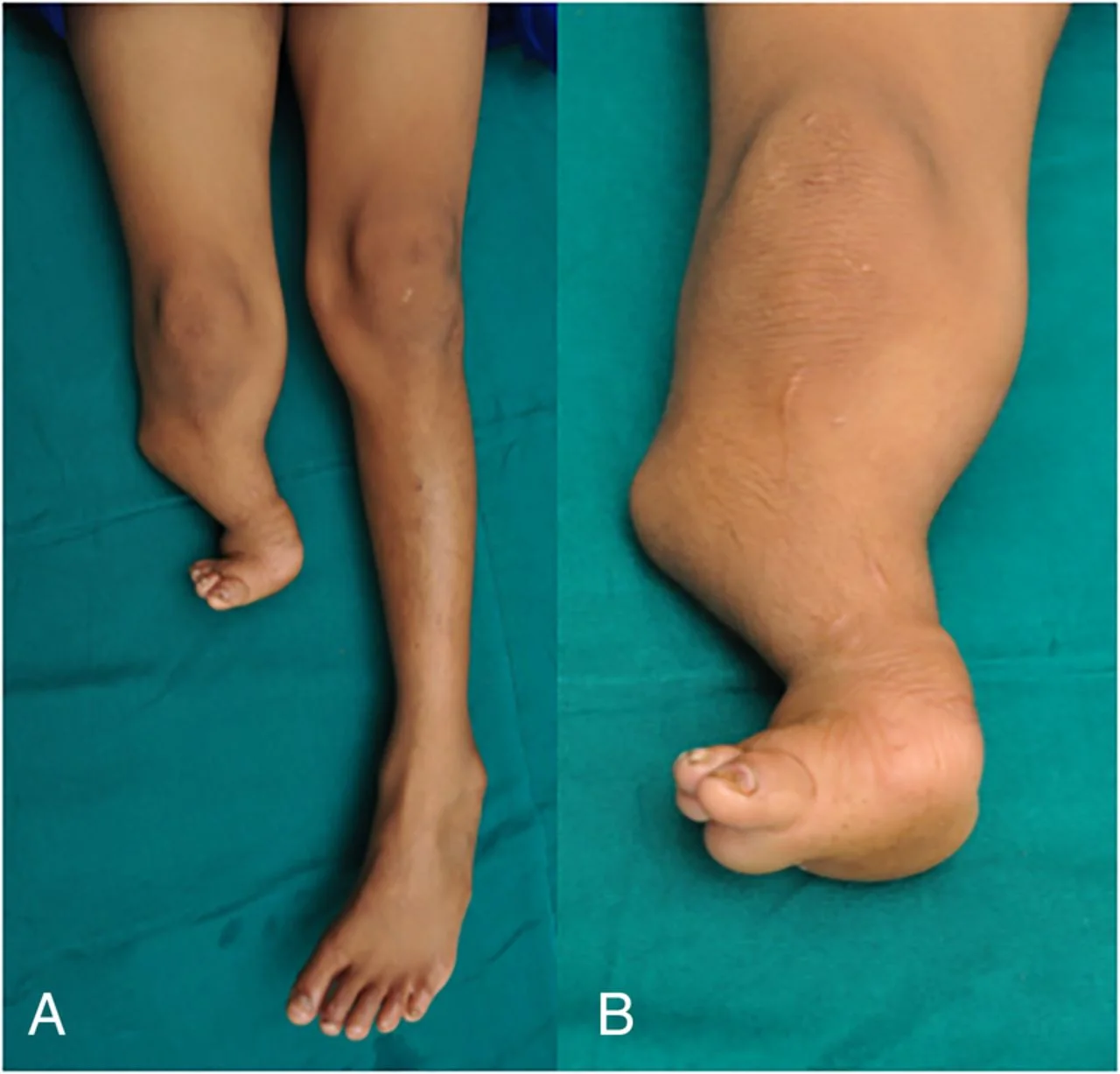
Tibial hemimelia is a rare congenital condition characterized by the partial or complete absence of the shinbone (tibia)—the main weight-bearing bone between the knee and ankle. In this complex anomaly, the tibia may be entirely absent (total) or only partially formed (partial).
Because the tibia carries the vast majority of the body's weight, its absence leaves the adjacent thinner bone (fibula) unable to support the leg properly. This results in significant leg shortening as well as severe deformities of the knee and ankle. It stands as one of the most complex and specialized congenital limb differences managed in pediatric orthopedics.
What are the symptoms?
Tibial hemimelia presents at birth with distinct, visually obvious signs:
Significant Leg Length Discrepancy: The affected leg is noticeably shorter than the healthy leg.
Knee Flexion Contracture: The knee joint cannot fully straighten and remains in a permanently bent (flexed) position.
Severe Foot and Ankle Deformity: The foot and ankle are severely turned inward, presenting a clubfoot-like appearance.
Lower Leg Instability: Absence of the tibia causes significant instability in the lower leg, often accompanied by deep skin folds.
Inability to Bear Weight: As the child reaches walking age, they are unable to stand or walk on the affected leg.
What causes it?
Tibial hemimelia stems from a developmental disruption during the early stages of fetal growth—specifically between the 4th and 7th weeks of pregnancy when the limb buds are forming.
While the exact cause remains unknown, it is occasionally associated with underlying genetic syndromes or familial traits. However, in the vast majority of cases, it occurs as an entirely isolated, random (idiopathic) developmental error with no prior family history.
How is it diagnosed?
Tibial hemimelia can be diagnosed both before and after birth:
Prenatal Diagnosis: Detailed high-resolution ultrasound scans performed during pregnancy can detect the absence or shortening of the leg bones while the baby is still in the womb.
Postnatal Diagnosis: Physical examination by a pediatric orthopedic specialist provides the initial diagnosis. Leg X-rays (Radiography) are then used to define the exact structural status, type, and severity of the deficiency. Additionally, Magnetic Resonance Imaging (MRI) is routinely performed to assess cartilage structures, knee ligaments, and surrounding muscles.
Treatment methods
The primary goal of treating tibial hemimelia is to enable the child to stand and walk on their own leg in a stable, pain-free manner. The treatment plan is fully individualized and surgically focused:
Reconstructive Surgery: In suitable cases where the bone deficiency is partial and knee ligaments are preserved, the adjacent fibula bone is repositioned to serve as the tibia (centralization), followed by leg lengthening techniques.
Prosthetic and Orthotic Support: Specialized pediatric prosthetic and orthotic systems are utilized throughout surgical management or post-operatively to support the leg and maximize function.
Is surgery required?
Yes. Surgical intervention is mandatory in all cases of tibial hemimelia, as there is no possibility of spontaneous self-correction without surgery.
If the shinbone is completely absent and the knee joint cannot be reconstructed, an early-age below-knee amputation (a limb-salvage approach) provides the best outcome, allowing the child to walk exceptionally well with a modern functional prosthesis. In partial deficiencies, a combination of procedures is performed—repositioning the fibula to act as the tibia, repairing joint ligaments, and utilizing Ilizarov or External Fixator systems for leg lengthening.
Recovery process
Managing tibial hemimelia is a long-term journey requiring patience and staged surgical follow-up. Following reconstruction and leg-lengthening procedures, bone consolidation and lengthening can take several months, during which external fixators remain attached to the leg.
After surgery, an intensive and continuous pediatric physical therapy program is crucial to preserve joint mobility and build muscle strength. In cases where amputation is chosen, the child adapts quickly to a functional prosthesis once the surgical wound heals, with the prosthetic length routinely adjusted as the child grows.
Frequently asked questions
Yes, children treated at the right time and with the right pediatric orthopedic strategy can walk, run and play sports independently. Children who undergo reconstructive surgeries gain mobility with their own legs, and children who undergo amputation gain mobility that is no different from their peers, thanks to modern carbon fiber prostheses.
Treatment planning should begin as soon as the baby is born. If leg lengthening and reconstruction surgeries are to be performed or surgical revision (amputation) is required for prosthesis compatibility, completing these procedures before the child reaches walking age (usually between 1-2 years of age) is the ideal time for the child's psychological and motor development.
Leg lengthening surgeries are very effective in suitable cases where the tibia is partially present and the knee/ankle joints are stable (intact). With the help of external fixators, the length of the leg is gradually lengthened to equalize it with the other leg. However, in severe cases where the tibia is completely destroyed, lengthening alone is not sufficient; it must be applied together with joint reconstruction.
Video
Appointment and information for Tibial Hemimelia
Contact us with your questions about the treatment process; we will arrange the most suitable appointment together.
 Çerez Tercihleri