



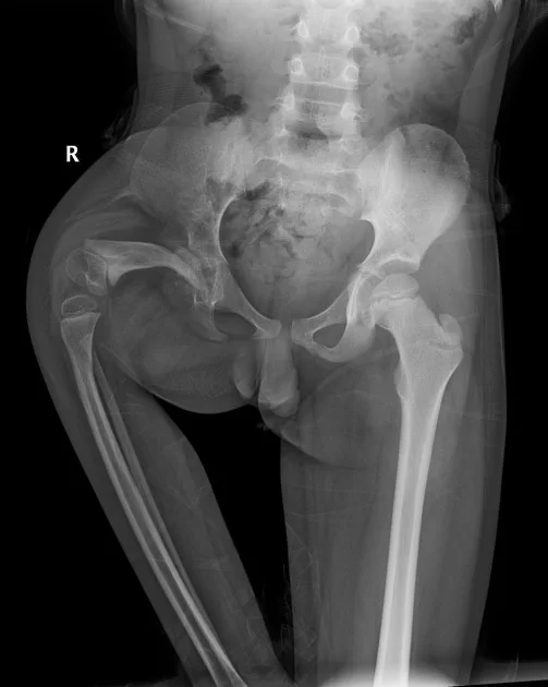
Proximal Femoral Focal Deficiency (PFFD)
Category Pediatric Orthopedics
Proximal Femoral Focal Deficiency is an anomaly characterized by congenital underdevelopment of the upper part of the femur, causing leg shortness and hip instability.
What is the problem?
Proximal Femoral Focal Deficiency (PFFD) is a rare congenital condition affecting the hip joint and the thighbone (femur). In this anatomical anomaly, the upper (proximal) portion of the femur near the hip either fails to develop entirely or experiences severely restricted growth.
As a result, the femur remains significantly shorter than normal, the hip joint loses its structural stability, and a major leg length discrepancy occurs. It stands as one of the most complex conditions in pediatric orthopedics, requiring a combination of advanced reconstructive surgery and specialized prosthetics.
What are the symptoms?
PFFD presents at birth with distinct visual characteristics that can be easily recognized:
Severe Thigh Shortening: The thigh area on the affected side is noticeably and significantly shorter than the other leg.
Abnormal Leg Posture: The shortened leg typically rests in an externally rotated (turned outward) and abducted (splayed out to the side) position.
Joint Instability: Marked laxity, instability, or restricted range of motion in the hip, knee, and ankle joints.
Muscle Underdevelopment: The musculature in the thigh region appears noticeably thinner and less developed.
Severe Gait Disturbance: As the child reaches walking age, the extreme leg length discrepancy prevents normal weight-bearing, resulting in a severe limp or an inability to stand on the affected side.
What causes it?
The exact cause of Proximal Femoral Focal Deficiency remains unknown. It arises from a developmental disruption or insult to the cells responsible for forming the thighbone during the very early stages of fetal development—specifically between the 4th and 6th weeks of pregnancy.
In most cases, PFFD is not inherited and shows no clear genetic link; it occurs as an entirely random (idiopathic) event during pregnancy.
How is it diagnosed?
Diagnosis relies on imaging techniques performed both before and after birth:
Prenatal Diagnosis (Antenatal Ultrasound): Detailed ultrasound scans performed during routine pregnancy checkups can detect the shortened thighbone (femur) at an early stage.
Postnatal Physical Examination: A pediatric orthopedic specialist clinically evaluates hip and knee stability as well as the exact leg length discrepancy.
X-Rays and MRI: Because a significant portion of an infant's skeleton consists of cartilage, Magnetic Resonance Imaging (MRI) is routinely performed to clearly visualize the hip socket (acetabulum) and femoral head. As the child grows, full-length standing X-rays (orthoroentgenograms) are used to measure the exact degree of ossification and bone length difference.
Treatment methods
The primary goal in treating PFFD is to ensure the child achieves independent, balanced, and functional mobility by the time they reach maturity. The treatment approach is divided into two main pathways based on the severity of the condition:
Limb Salvage and Leg Lengthening: In milder cases where the femoral deficiency is limited and the hip joint can be stabilized, multi-stage bone lengthening procedures are performed.
Surgical Reconstruction for Functional Prosthetics: In severe cases where bone loss is extensive, specialized limb-salvage surgeries are performed to align the limb biomechanically and optimize it for comfortable, highly functional prosthesis use.
Is surgery required?
Yes. Surgical intervention or surgically enabled prosthetic rehabilitation is mandatory in all cases of PFFD.
In milder cases, bone-reshaping procedures (osteotomies) are performed to stabilize the hip joint, followed by leg lengthening surgeries using external fixators. In severe cases, two major world-standard surgical procedures are widely utilized:
Hip Stabilization (e.g., King Procedure): The femur and pelvis are reconfigured to improve the weight-bearing capacity and stability of the hip joint.
Van Nes Rotationplasty: In cases where the knee joint is non-functional, the lower leg and foot are rotated 180 degrees so that the ankle joint functions as a knee joint. This allows the child to fit a below-knee prosthesis rather than an above-knee prosthesis, resulting in significantly better mobility and functional outcomes.
Recovery process
PFFD management is a long-term, multi-stage treatment journey requiring immense patience that continues until the child completes their growth period (late adolescence).
If lengthening procedures are chosen, each phase involves months of external fixator wear and ongoing clinical follow-up while the new bone consolidates. In cases involving limb modification surgeries, such as Rotationplasty or amputation, the child begins prosthetic training shortly after wound healing—typically within 6 to 8 weeks. Throughout every stage of treatment, continuous pediatric physical rehabilitation is essential to prevent joint stiffness and maintain muscle strength.
Frequently asked questions
Yes, thanks to today's modern pediatric orthopedic surgery and advanced prosthetic technologies, children with PFFD can walk, run, go to school and lead active lives independently. Success in treatment allows the child to catch up with his peers with his own leg or a high-functioning prosthesis.
In severe cases of PFFD, the knee joint cannot function because the thigh is too short. With rotationplasty surgery, the child's own ankle is turned upside down and brought to knee level. Thus, the ankle joint begins to function like a new "knee joint". In this way, the child can run and play like his peers with a comfortable below-knee prosthesis that he can control with his own muscles, instead of an above-knee prosthesis that is very difficult to use.
Leg lengthening surgeries alone are sufficient only in mild forms of PFFD (Type A and partially Type B according to the Aitken Classification) and if the structure of the hip/knee joints can be corrected early with surgery. In severe cases where the leg length difference is more than 50%, lengthening alone is not biomechanically possible; in these cases, prosthetic-oriented surgeries should be preferred.
Video
Appointment and information for Proximal Femoral Focal Deficiency (PFFD)
Contact us with your questions about the treatment process; we will arrange the most suitable appointment together.
 Çerez Tercihleri