


scoliosis
Category Orthopedics
Scoliosis is a three-dimensional curvature of the spine greater than 10 degrees to the right or left. With early diagnosis, it can be treated with corset and in advanced stages with surgery.
What is the problem?
Scoliosis is defined as a lateral curvature of the spine of 10 degrees or more to the right or left, occurring in the thoracic (chest) or lumbar (lower back) regions. However, scoliosis is not merely a two-dimensional curve; it is a complex, three-dimensional spinal deformity that also involves the rotation of the vertebrae along their axis.
Most commonly emerging just before the adolescent growth spurt, this condition—if left untreated—can lead to severe limitations in pulmonary and cardiac function alongside significant cosmetic concerns. In spinal health and orthopedics, early diagnosis plays a critical role in halting progression.
What are the symptoms?
Scoliosis rarely causes pain in its early stages. Therefore, parents should remain vigilant for the following physical signs, particularly during childhood and adolescence:
Uneven Shoulder Heights: One shoulder appearing higher or more prominent than the other.
Asymmetrical Hips: One side of the hip appearing higher, protruding, or uneven.
Rib Hump or Prominence: A visible bulge or hump on one side of the back when the child bends forward (the Adam's Forward Bend Test position).
Asymmetrical Arm-to-Trunk Gap: An uneven space between the hanging arms and the sides of the torso.
Prominent Shoulder Blade: One scapula sticking out or standing out more prominently than the other.
What causes it?
The causes of scoliosis are classified into distinct categories based on their structural characteristics:
Idiopathic Scoliosis: Accounting for approximately 80% of all scoliosis cases, its exact cause remains unknown, though genetic predisposition is believed to play a significant role.
Congenital Scoliosis: Present at birth, this type develops during fetal development in the womb due to malformed or incompletely formed vertebrae.
Neuromuscular Scoliosis: Arising secondary to neuromuscular disorders such as Cerebral Palsy, muscular dystrophy, or polio, where impaired muscle control prevents adequate support of the spine.
How is it diagnosed?
Adam's Forward Bend Test: During the physical examination, the patient is asked to bend forward, and any back asymmetry (rib hump) is measured using a scoliometer.
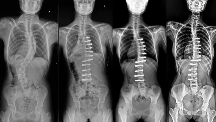
Full-Spine Radiography (Scoliosis X-Ray): Standing anteroposterior and lateral X-rays are taken to calculate the exact degree of spinal curvature (Cobb Angle).
Magnetic Resonance Imaging (MRI): Requested to evaluate the spinal cord structure in cases of rapidly progressing curves, congenital conditions, or when neurological involvement is suspected.
Treatment methods
Scoliosis treatment is personalized based on the severity of the curve (Cobb angle), the patient's age, and their remaining growth potential:
Observation and Monitoring: For curves under 20 degrees in patients approaching the end of their growth period, regular follow-ups every 4 to 6 months are conducted to monitor for potential progression.
Scoliosis Exercises: Specialized physiotherapy techniques (such as the Schroth Method) help balance muscle strength and support the overall treatment plan.
Bracing Therapy: For growing children with curves between 20 and 40 degrees, custom-fitted braces worn 16 to 23 hours a day are utilized to prevent the curvature from progressing.
Is surgery required?
Yes. Surgical intervention is required for growing children whose curves continue to progress despite bracing therapy, or for those whose initial diagnosis reveals a curve exceeding 40 to 45 degrees.
In adulthood, curves greater than 50 degrees can continue to advance due to the force of gravity and may compromise lung and heart function; therefore, surgical correction is recommended in these cases as well.
Recovery process
In modern spinal fusion (posterior fusion) procedures, the spine is realigned and stabilized using titanium screws and rods. Patients are typically mobilized and walking by the first or second post-operative day, with an average hospital stay of 4 to 5 days.
Thanks to advanced surgical techniques and rigid internal fixation, post-operative casts or braces are rarely required. Students can generally return to school within 3 to 4 weeks. Full bony fusion and a gradual return to active sports and physical activities (excluding high-impact contact sports) are typically achieved within a 6- to 12-month follow-up period.
Frequently asked questions
Swimming or general sports activities strengthen the back muscles, reduce pain and improve posture, but they alone do not correct the structural curvature (Cobb angle) in the bone. In cases of scoliosis with a high degree and risk of progression, evidence-based special scoliosis exercises (Schroth method) and brace treatment should be applied under the supervision of a pediatric orthopedist.
The primary purpose of brace treatment is to prevent further progression of the curvature in the spine during the child's rapid growth period and to protect the patient from reaching the surgery stage. If the corset is used regularly for the period recommended by the doctor (usually around 20 hours a day), the success rate is quite high and in some cases, permanent improvements in the degree of curvature can be observed.
No, on the contrary, since the curves in the spine are corrected during scoliosis surgery, a sudden increase of a few centimeters in the patient's height is observed immediately after the surgery. Although the lengthening of the vertebrae in the area fixed (fused) during surgery stops, the natural growth and height of the child continues in its normal course, thanks to the leg length and other non-fixed spinal areas.
Video
Appointment and information for scoliosis
Contact us with your questions about the treatment process; we will arrange the most suitable appointment together.
 Çerez Tercihleri